
Products
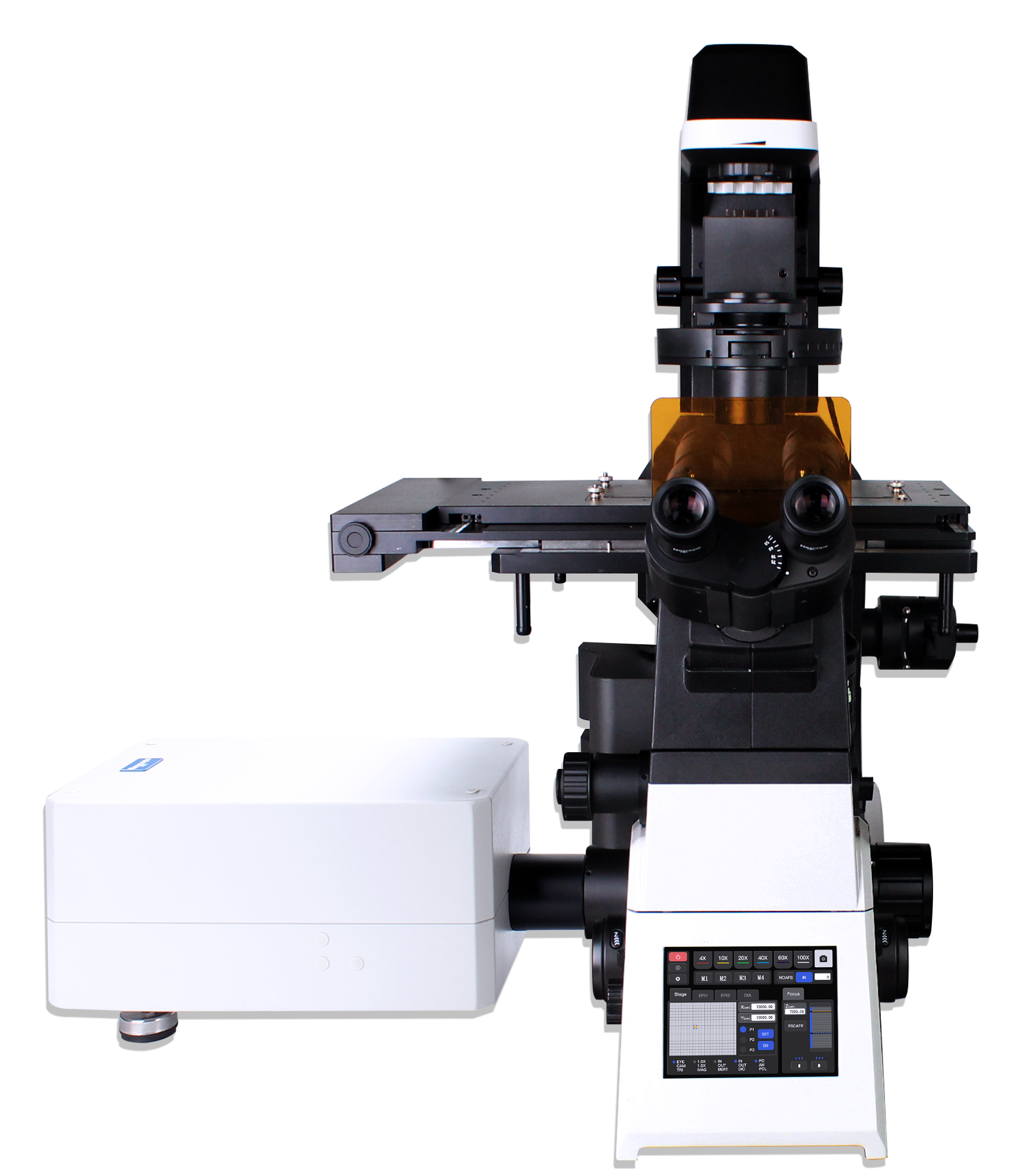
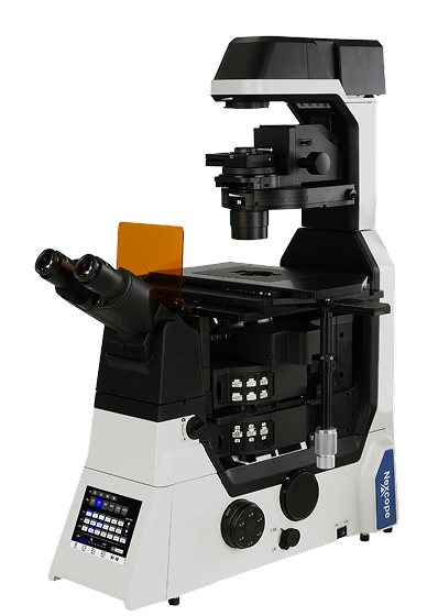
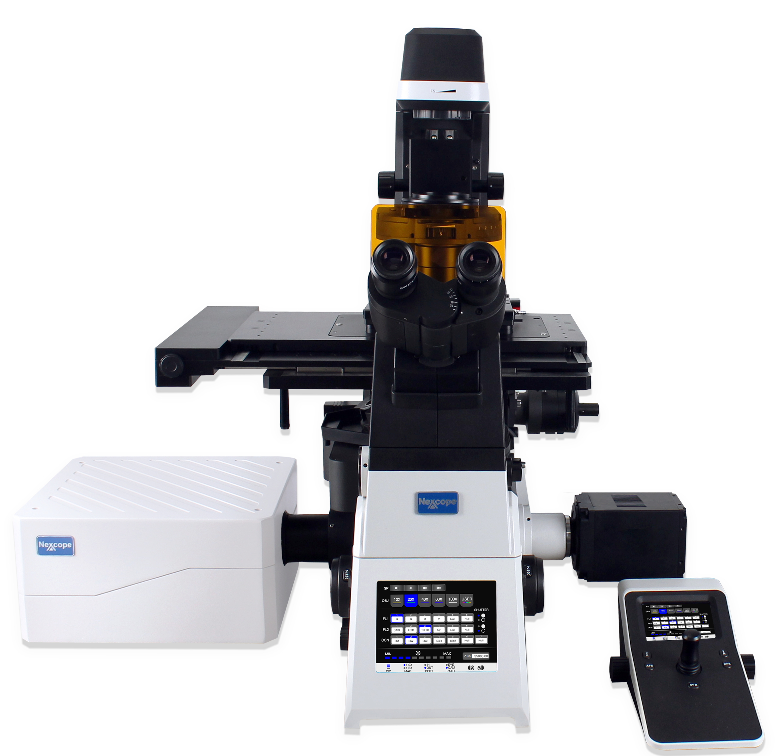
As life‑science research advances toward deeper dimensions and broader scopes, high‑efficiency imaging technology has become a core driver for scientific breakthroughs. Capturing images of large‑size specimens requires not only expanded imaging coverage but also drastically shortened acquisition time, so as to precisely record intracellular dynamic responses. A full‑scale upgrade over the NCF1000, the NCF2000 laser confocal microscope is built to meet the extreme demands for high‑throughput, high‑quality imaging in modern biological research.
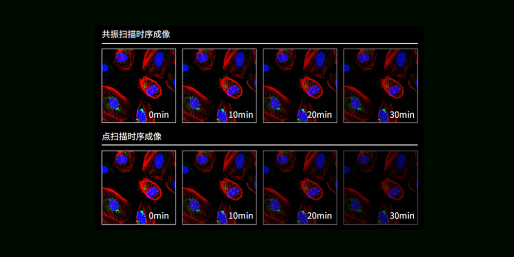
High‑speed Resonant Scanning: Resonant‑scan technology delivers ~20× faster speed, lowering photodamage for long‑term live‑cell observation.
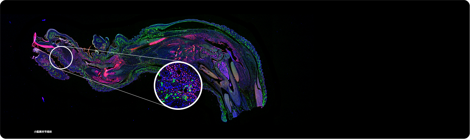
Fast High‑definition Imaging: Supports rapid 8K×8K imaging; premium optics capture subtle specimen details.
Gentler Imaging for Live‑cell Samples: Reduced laser exposure preserves cell viability, enabling fast dynamic recording and long‑term time‑lapse for drug and microplate screening.

High‑speed Resonant Scanning: Resonant‑scan technology delivers ~20× faster speed, lowering photodamage for long‑term live‑cell observation.
Fast High‑definition Imaging: Supports rapid 8K×8K imaging; premium optics capture subtle specimen details.
Gentler Imaging for Live‑cell Samples: Reduced laser exposure preserves cell viability, enabling fast dynamic recording and long‑term time‑lapse for drug and microplate screening.

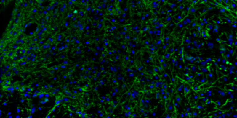
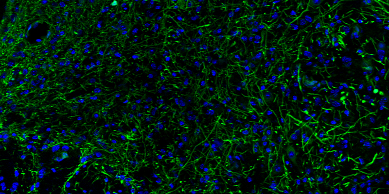
For images of equivalent quality, the illumination exposure time is reduced, which mitigates photobleaching and phototoxicity to samples. This facilitates long‑term observation of sensitive specimens such as live cells.

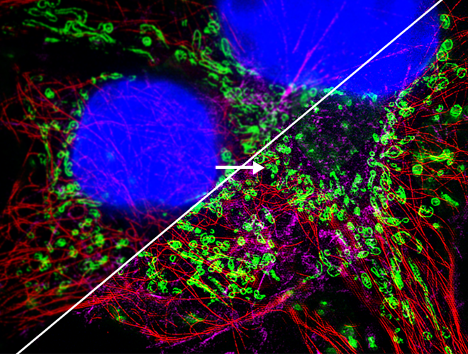
Conventional Detector Imaging
Limited capability for capturing faint fluorescence signals, often resulting in weak signals, high noise and blurry outputs. Low resolution makes it difficult to clearly resolve fine microstructures; tiny features are easily lost, failing to satisfy imaging requirements for weak‑signal and high‑detail samples.

Array‑based Detector Imaging
Accurately captures extremely faint fluorescence signals for crisp, detail‑rich imaging. High resolution resolves specimen microstructures with fine granularity, rendering minute features clearly visible.

High Fluorescence Collection Efficiency
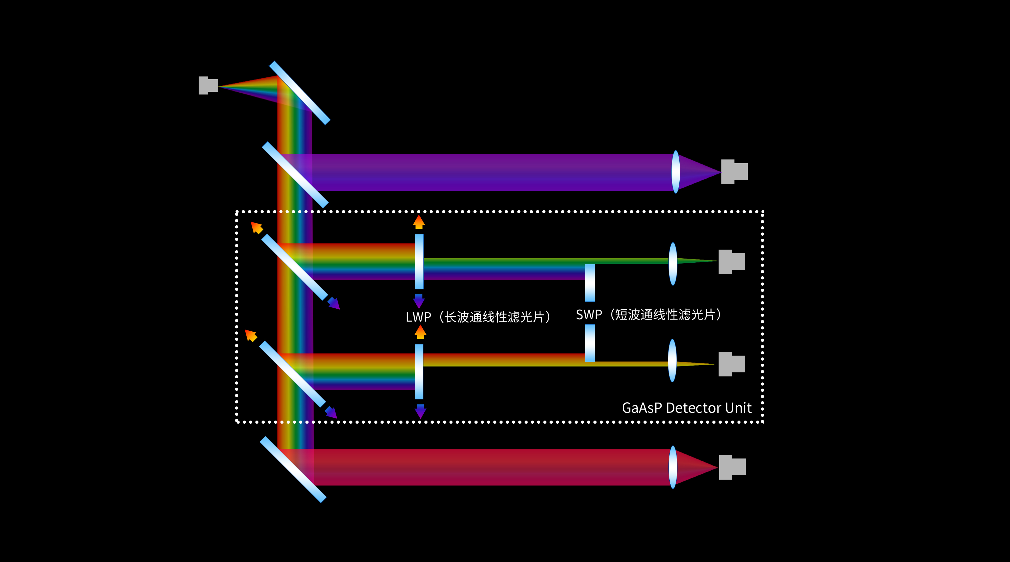
Equipped with GaAsP photomultiplier tubes (PMTs), the NCF2000 improves fluorescence collection efficiency, allowing observation of faint‑fluorescence samples that are hard to detect with conventional methods. The system supports up to four distinct spectral channels for effective spectral decomposition of sample‑emitted fluorescence. It achieves spectral resolution ≤ 2 nm, suppresses background noise under low‑excitation‑light conditions, and delivers outstanding multi‑color confocal imaging performance.

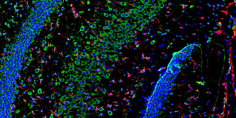
Spectral‑unmixing Function
It performs spectral decomposition on fluorescence emitted from samples to obtain fluorescence images and spectral information at different wavelengths. This enables differentiation of substances with distinct fluorescence‑emission profiles, even when they appear visually similar under conventional light microscopes. Spectral‑feature analysis supports identification and characterization of specific chemical components or biomolecules within specimens.

Nomis Pro X‑C
High‑speed Hardware Control: Provides streamlined digital management and precise control of multiple motorized components inside the microscope.
Multi‑dimensional Imaging & Image Display: Supports combined X, Y, Z, λ, T scan modes, with multiple flexible built‑in shooting presets.
Deconvolution: Performs deblurring for 2D images. Multiple rounds of deconvolution can be applied to eliminate shot‑noise components in confocal images.

Nomis Pro X‑C
High‑speed Hardware Control: Provides streamlined digital management and precise control of multiple motorized components inside the microscope.
Multi‑dimensional Imaging & Image Display: Supports combined X, Y, Z, λ, T scan modes, with multiple flexible built‑in shooting presets.
Deconvolution: Performs deblurring for 2D images. Multiple rounds of deconvolution can be applied to eliminate shot‑noise components in confocal images.

Observation modules
User‑friendly controls and seamless modality switching to build an efficient imaging environment for researchers.

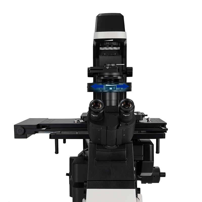
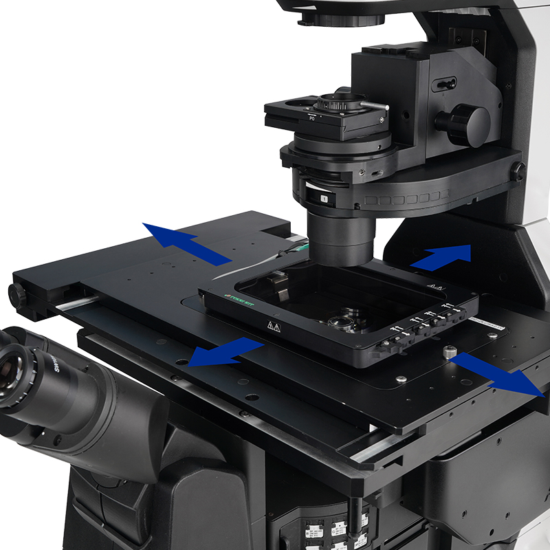
XY Motorized Stage:
Responsive travel and stable positioning, paired with intuitive joystick control for effortless operation.

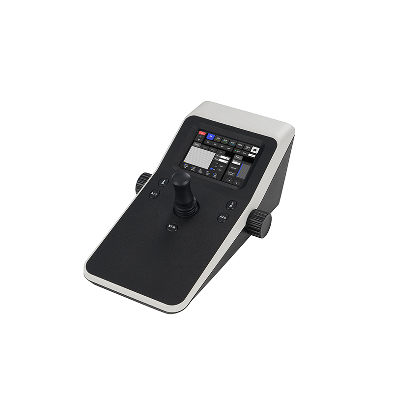
Joystick with Built‑in Touch Panel
Equipped with an integrated touch screen, the joystick allows configuration of instrument status, stepped control for the motorized stage and motorized focusing. It also features dedicated function keys and an AFS button.

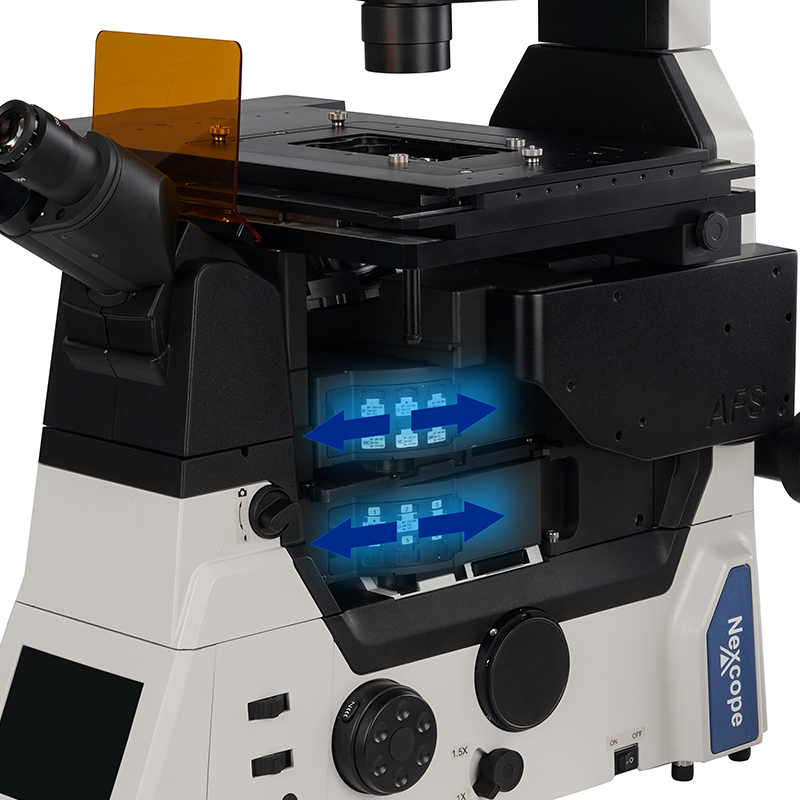
Filter blocks
Fast switching and highly‑accurate positioning, enabling rapid transitions between different observation modes.

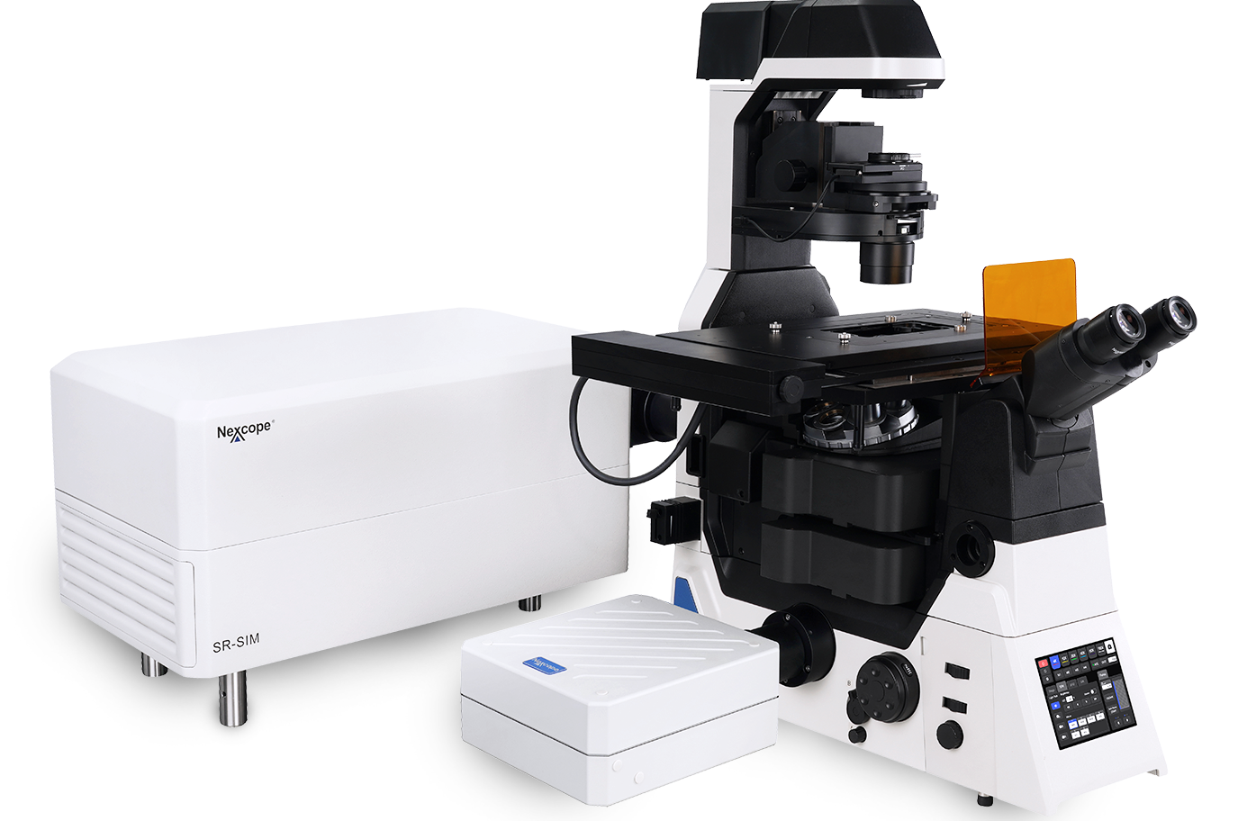
Super‑Resolution Module

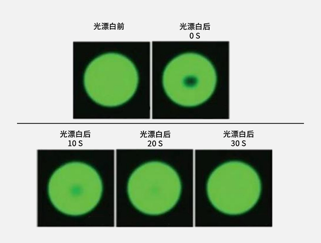
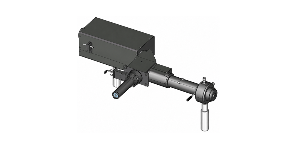
FRAP (Fluorescence Recovery After Photobleaching) Module

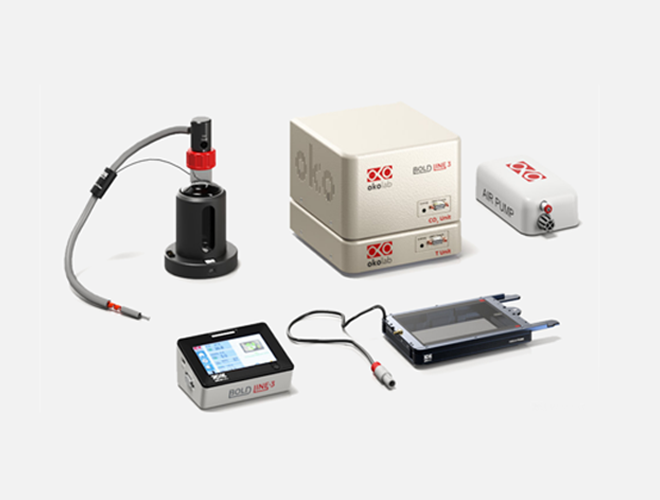
Live‑Cell Incubation System

TIRF Module

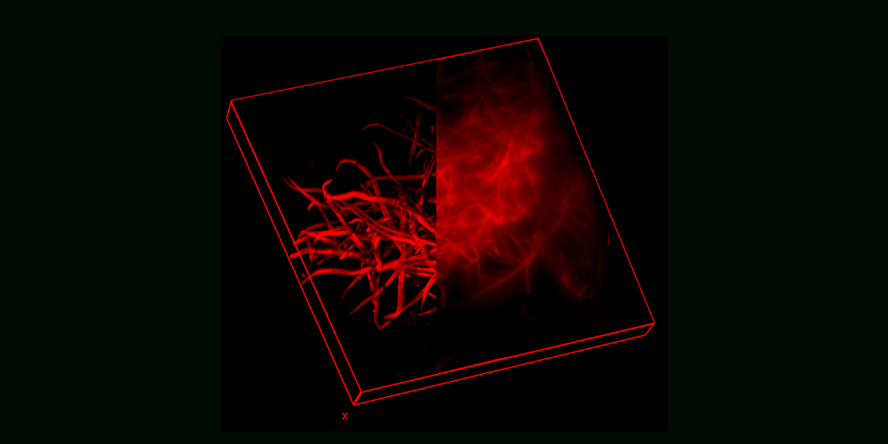
Optical Sectioning Module