
 Products







First, experimental background: microneedle patch
1. Microneedles (MN) is a new physical penetration-promoting technology, which consists of a plurality of micron-sized fine needle tips connected to a base in an array. The needle body is generally 10-2,000 microns high and 10-50 microns wide. The length, size and shape of microneedles can be individually designed according to the needs of treatment. Micro-needles can directionally pass through the stratum corneum, producing micron-sized mechanical channels, placing drugs directly on the epidermis or upper dermis, and participating in microcirculation without passing through the stratum corneum to exert pharmacological reactions.
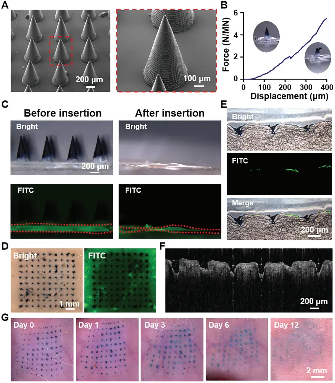
Figure 1 is quoted from: Small. 2025 Jan; 21(3):e2409121.
2. When microneedles deliver drugs, the active ingredients are loaded in the microneedle array, and the concentration gradient between drugs and subcutaneous tissue fluid forms the driving force, so that drugs are slowly released into the body. Besides the advantages of transdermal delivery system, it also has the following advantages:
(1) the transdermal absorption rate is stable;
(2) Controlling the length of microneedles can avoid touching capillaries and nerve endings and reduce or eliminate pain;
(3) The administration mode is convenient and can be used by oneself.
Second, the experimental scene: Z-stack-3D reconstruction
1. Z-stack(Z-layer stacking and Z-axis sequential scanning) is a three-dimensional imaging acquisition technology of confocal laser scanning microscope: along the Z-axis of the sample (perpendicular to the axial direction of the slide/objective lens and the depth direction), a plurality of two-dimensional confocal slice images with different focal planes are acquired layer by layer at a fixed step (a single slice is called Z-slice), and a set of continuous depth slices is called Z-stack stack.
Step 2: Use steps
Z-axis slice scanning: the shooting mode can manually set the starting coordinates and ending coordinates, flexibly frame the slice scanning range, and set the Z-axis step size or scanning steps during scanning. Note: the smaller the step size, the larger the scanning steps, the finer the scanning and the better the effect.
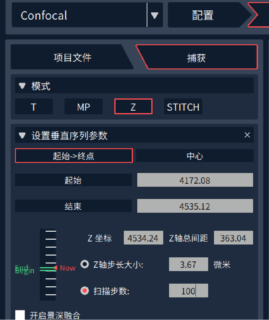
Figure 2: Layer Scan Settings
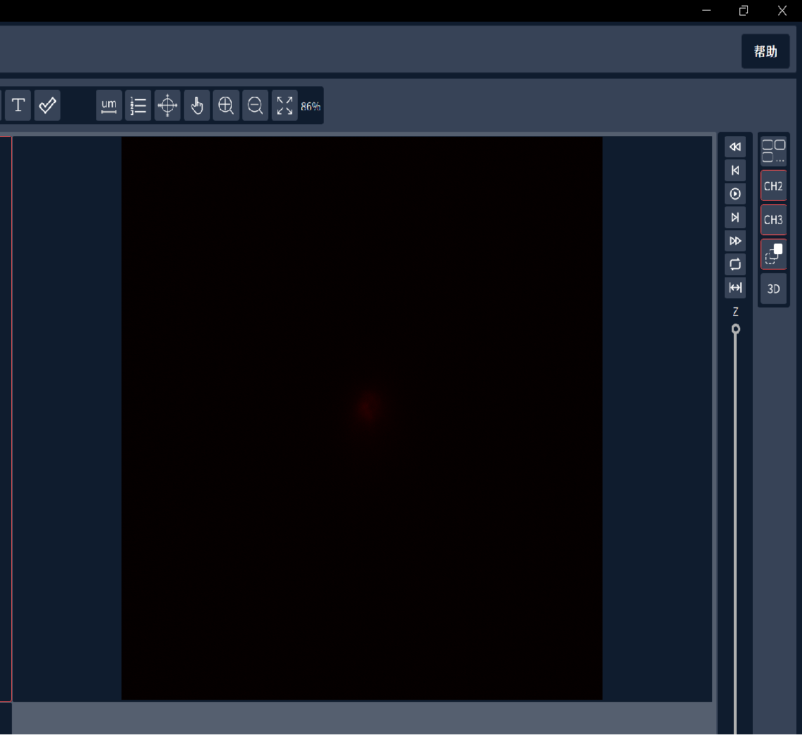
Figure 3: 3D reconstruction
3D reconstruction: After Z-axis slice scanning, a "3D" button will appear in the software. Click this button to enter the new interface and automatically reconstruct the photographed sample.
Third, the observation effect of the user's microneedle patch
In this experiment, the user dyed the microneedle carrier and the drug separately. The drug was excited by a 488nm laser and the carrier was excited by a 561nm laser. First, the overall situation of the microneedle was observed by using a 10X objective lens, and then switched to a 20X objective lens for Z-stack scanning and 3D reconstruction.
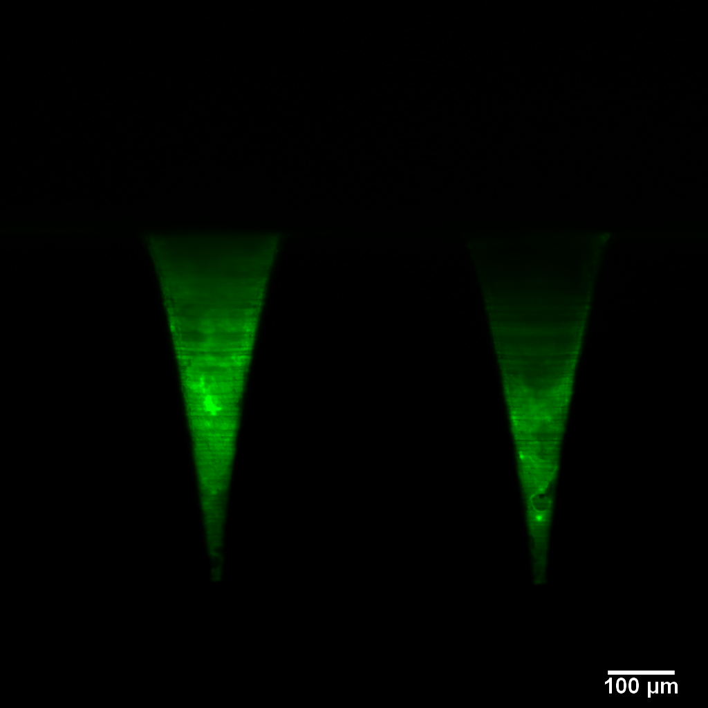
Figure 4: Drugs
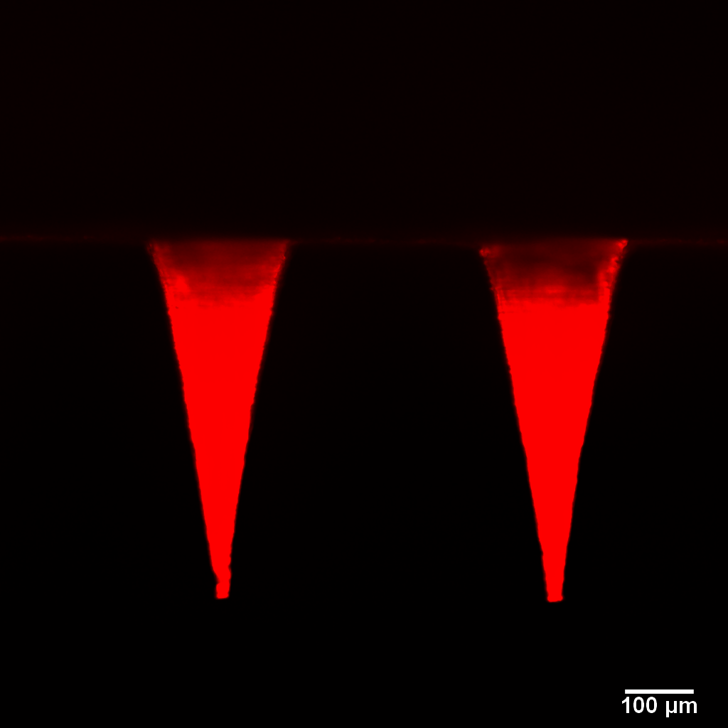
Figure 5: Carrier
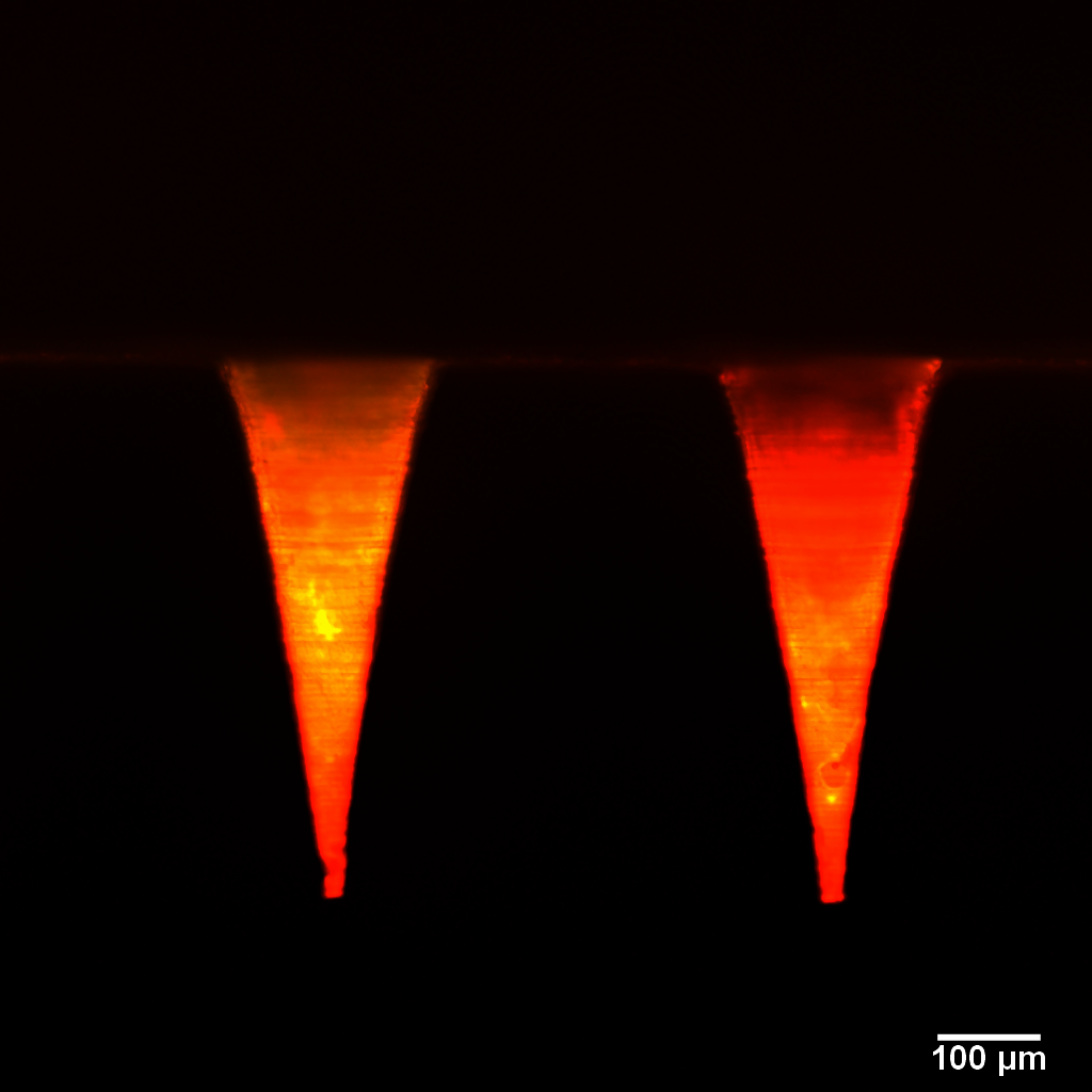
Figure 6: Merge diagram
In this experiment, 3D reconstructs schematic diagrams from different perspectives. The green highlighted part in the picture is the drug aggregate carried.
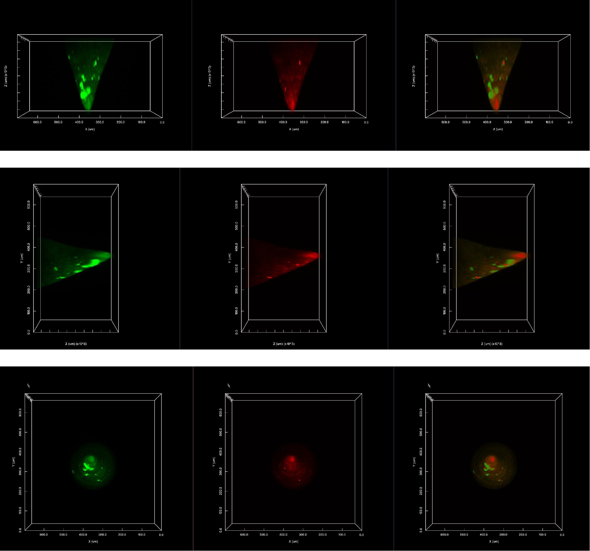
Z-stack shooting conditions
Microscope: NCF1000 laser confocal microscope.
Objective lens: 20X (NA = 0.75)
Resolution: 1024 X 1024 pixels
Laser channel: 488nm, 561nm.
Number of layer scanning steps: 120
Single picture shooting condition
Microscope: NCF1000 laser confocal microscope.
Objective lens: 10X(N.A.=0.45)
Resolution: 2048 X 2048 pixels
Laser channel: 488nm, 561nm.
Fourth, the problems that may be encountered during the experiment
Q1: When using gel microneedles, what should I do if they melt and become soft?
A1: When the state of the microneedle changes during shooting, you can put the microneedle on the cover glass and observe it upside down by virtue of its own viscosity.