
 Products







1. Principle and application scenarios of reflective confocal.
1. Imaging principle: Reflective confocal microscope uses laser scanning imaging, and generates back reflection signal depending on the refractive index difference inside the sample. After the reflected light returns, the confocal pinhole can filter out defocused stray light and only collect the effective information of the focal plane. The system scans point by point and reconstructs the image by computer. Scanning layer by layer can realize three-dimensional optical tomography. This technique can be used for noninvasive observation of living tissues without fluorescent labeling.
2. Application scenarios: Reflective confocal microscopy imaging has rich application scenarios with the characteristics of label-free and non-invasive optical tomography. In clinical medicine, this technique is most commonly used in dermatology, and it can be used for in vivo screening and curative effect follow-up of pigmented nevus, melanoma, basal cell carcinoma and other lesions without tissue biopsy. At the same time, it is also used in ophthalmology to realize in vivo observation of corneal epithelium and matrix collagen, and to assist in evaluating corneal lesions and transplant conditions. The field of scientific research can be used to observe the microstructure of mucosal tissue and tissue engineering scaffold. In material and industrial inspection, it can detect surface and subsurface defects, interface morphology and coating structure of films, composites and optical components. Limited by imaging depth, this method is suitable for rapid and nondestructive evaluation of surface microstructure.
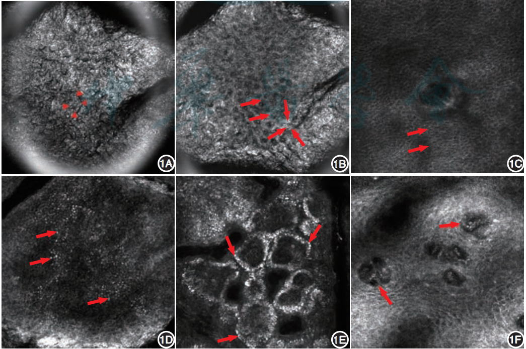
Fig. 1: Reflective confocal skin tissue imaging pictures from: Chin J Dermatol, September 2024, Vol.57, No.9.
Second, use NCF1000 reflective confocal to observe the crystal structure.
1. Crystal surface imaging
In this experiment, the user uses a 20X 0.75 objective lens to perform reflective confocal imaging on crystal fragments. The software is switched to reflective confocal mode, and the appropriate wavelength laser is selected according to the experimental materials. NCF1000 is equipped with lasers of 405 nm, 488 nm, 561 nm and 640 nm. When the numerical aperture of the objective lens and the refractive index of the medium remain the same, the optical layer thickness increases with the increase of laser wavelength. The thinnest optical slice can be achieved at 405 nm with the best axial resolution, but the tissue light scattering is strong and the penetration depth is limited. With the wavelength increasing to 488 nm and 561 nm, the lamellar thickness gradually increases and the tissue penetration ability is improved. The slice thickness of 640 nm is the largest and the axial resolution is the weakest among the four, but it can effectively reduce the scattering of biological tissues and obtain deeper imaging. When choosing laser, we need to weigh the axial slice accuracy, fluorescent probe matching and tissue penetration depth. Adjust the appropriate laser and gain intensity, and it is recommended to switch the aperture size to 1.5-2AU.

Figure 2: Crystal surface imaging
Shooting conditions:
Microscope: NCF1000 reflective confocal microscope.
Objective lens: 20xna: 0.75
Resolution: 2048 X 2048 pixels
Laser: 488nm pinhole: 1.5AU
2. Crystal 3D structure imaging
NCF1000 reflective confocal can also shoot Z-Stack, and use Nomis Pro X_C software for three-dimensional reconstruction.
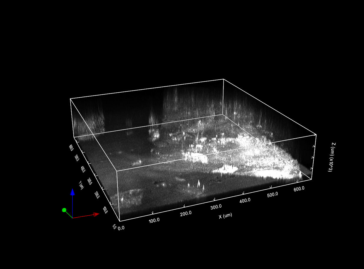
Figure 3: 3D structure imaging of crystal
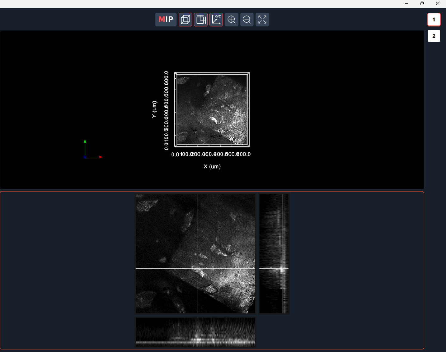
Figure 4: Slicing mode can be used to observe slicing signals at different positions.
Fourth, the problems that may be encountered during the experiment
Q1: When imaging in Z-stack mode, can the saved original file be saved in a folder?
A1: At present, Z-Stack scanning original files saved by Nomis Pro X_C software are not supported to be saved in a folder for the time being.
Note: When performing reflective confocal imaging on the crystal, it is necessary to pay attention to the distance between the sample and the objective lens. Prevent the crystal from scratching the objective lens.