I. Experimental Scenario: Observation of Lymphoma Cells
1.1 Pfeiffer Lymphoma Cells
Lymphoma refers to a group of malignant tumors originating from the lymphatic system, featuring high heterogeneity and complexity. In‑depth research on the biological properties of lymphoma cells is irreplaceably important for basic medicine, clinical therapy and public health. Pfeiffer cells represent a subtype of Diffuse Large B‑Cell Lymphoma (DLBCL), with molecular characteristics distinct from classic DLBCL. Targeted therapy designs drugs against unique molecular markers of Pfeiffer cells, such as specific gene mutations or abnormal protein expression, to achieve precise tumor suppression, reduce damage to normal cells and improve therapeutic efficacy.
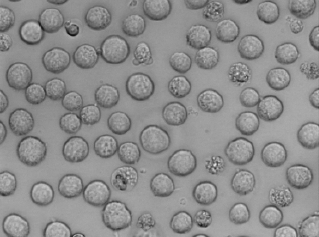
Figure 1 Pfeiffer: Revived human diffuse large B‑cell lymphoma cells
1.2 Application of Confocal Microscopy on Pfeiffer Lymphoma Cells
- High Resolution and 3D Imaging
Observation of subcellular structures: Surpassing conventional light microscopes in resolution, confocal microscopes enable clear visualization of subcellular structures of Pfeiffer cells including nuclei, mitochondria and endoplasmic reticulum. For instance, fluorescence labeling allows precise localization of intracellular DNA or RNA within nuclei, revealing genetic abnormalities in lymphoma cells.
3D reconstruction: By scanning samples layer‑by‑layer and stacking optical sections, confocal microscopes reconstruct three‑dimensional architectures of Pfeiffer cells. Intuitive visualization of cellular morphology and spatial distribution facilitates understanding of tumor‑cell heterogeneity and growth patterns.
- Fluorescence Labeling and Molecular Localization
Fluorescent probes such as antibodies and nucleic acid dyes label specific molecules in Pfeiffer cells (e.g., tumor‑associated antigens, signaling‑pathway proteins) for detection via confocal microscopy.
- Drug Development and Mechanism Research
Drug action mechanism: Confocal microscopes characterize drug effects on Pfeiffer cells, including drug‑induced apoptosis and autophagy. For example, changes in mitochondrial membrane potential in drug‑treated Pfeiffer cells can be monitored to evaluate pro‑apoptotic drug effects.
1.3 Observation Objectives
To explore the pathogenesis of lymphoma and potential targeted therapeutic regimens, researchers studied HMGA2 and CDCA8. Under confocal microscopy, changes in protein fluorescence intensity after gene knockout were to be observed.
1.4 Imaging of Lymphoma Cells
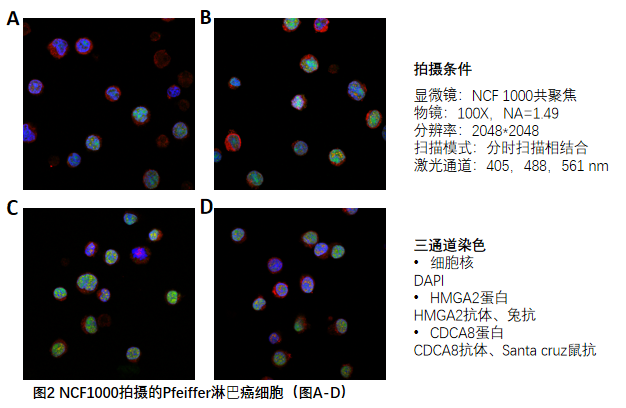
In this experiment, DAPI was used for nuclear visualization. Anti‑HMGA2 and anti‑CDCA8 antibodies served as primary antibodies. Rabbit secondary antibody and Santa Cruz mouse secondary antibody were applied for labeling HMGA2 and CDCA8 respectively. Equipped with a 100×, 1.49 NA high‑magnification apochromatic objective, the NCF1000 confocal microscope imaged Pfeiffer cells under 405 nm, 488 nm and 561 nm excitation channels (Figure 2 A‑D).
II. How NCF1000 Confocal Microscope Supports the Experiment
2.1 Image‑Centering Function
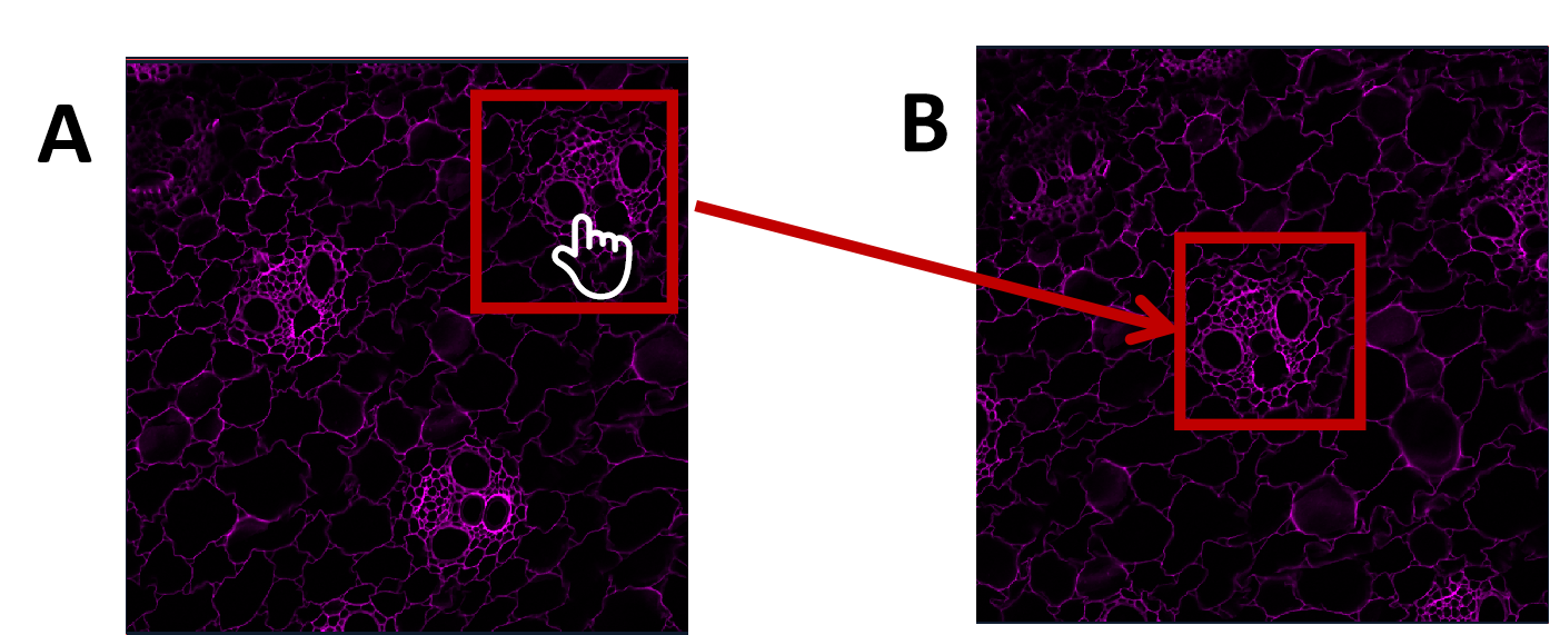
Image‑centering quickly moves targets to the center of the field of view. With the hand‑tool selected, double‑click the target to shift it to the field‑of‑view center.
(B)Figure 3 Image‑centering function: Double‑click target object (A); target moved to field‑of‑view center (B)
2.2 Multi‑Site Scanning
Multi‑site scanning enables acquisition of multiple fields‑of‑view on one sample slide under identical experimental conditions including laser intensity, gain, pinhole size and resolution. It improves operational convenience and supports direct multi‑region comparison.
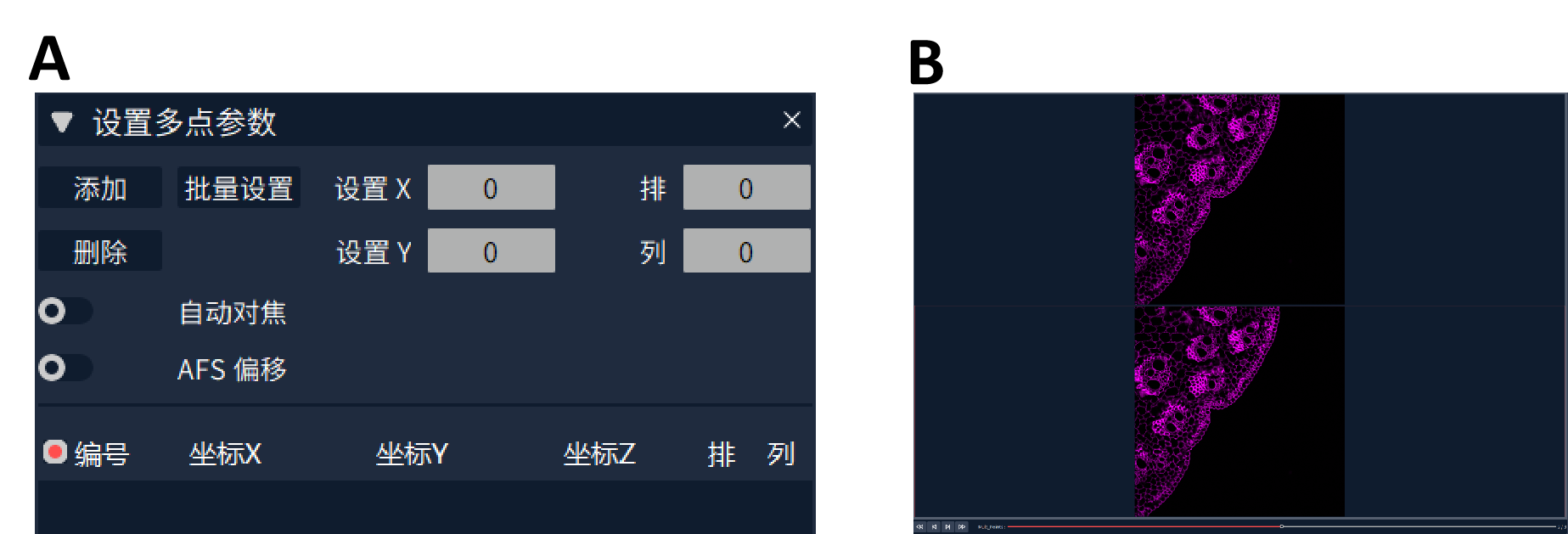
Two field‑of‑view setting modes are available: custom‑defined positions and automatic position generation. Multi‑site scanning stores X‑, Y‑ and Z‑axis coordinates of each field‑of‑view center. For scattered individual cells, multi‑site scanning can be combined with ROI cropping for single‑cell‑oriented multi‑position acquisition. After adding positions, double‑click coordinate entries to drive stage movement for field‑of‑view re‑verification.
Figure 4 Multi‑site scanning interface and image preview: Multi‑site scanning mode (A); preview targets at different positions via image playback (B)
2.3 Reuse of Image Parameters
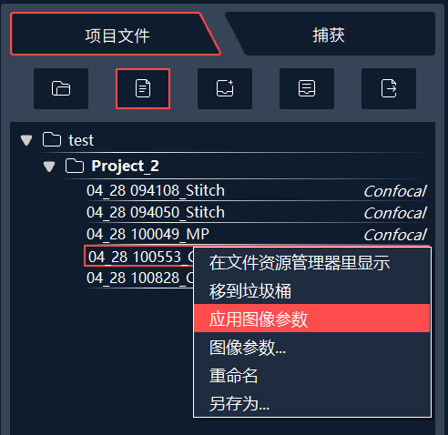
For samples requiring imaging under identical settings, reuse existing image parameters to guarantee consistent experimental conditions. Select the reference image, right‑click and choose “Apply Image Parameters” (Figure 6). Channel, pinhole, position, laser power, gain and other parameters will be loaded to software and hardware.
Figure 6 Operation for applying image parameters
III. Common Experimental Issues
Q1: How to rapidly export multiple captured image sets?
A1: Under the capture folder, select target files, right‑click and select batch image export. Customize storage path and file names before confirming export. Each dataset generates an independent folder containing single‑channel and merged‑channel images.
Q2: Few cells on sample; targets are hard to locate under high‑magnification objectives in confocal mode. How to find specimens efficiently?
A2: Locate samples under wide‑field mode. The wide‑field mode delivers a larger field‑of‑view for fast target identification. After locating targets, switch software to confocal mode. Confirm left‑side optical path and empty fluorescence module before confocal acquisition.

 Products