I. Experimental Scenario: Mitochondrial Membrane Potential Detection of MDA‑MB‑231 Cells
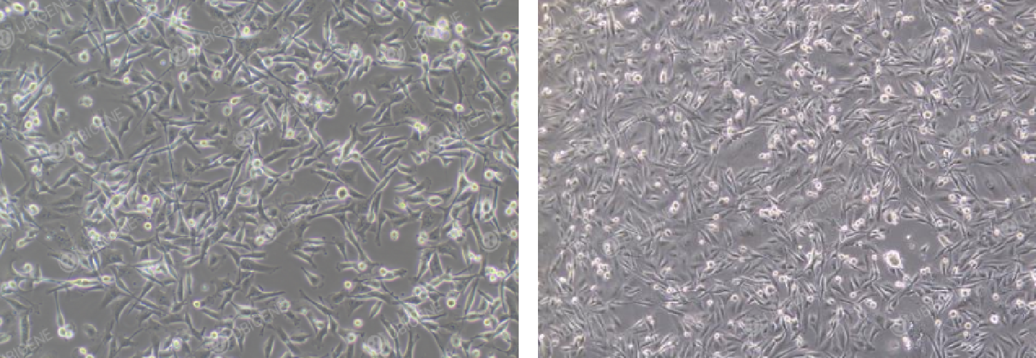
1. MDA‑MB‑231 cells (human breast cancer cells) are metastatic human breast cancer cells widely used in cancer research. As epithelial cells, most cells exhibit spindle‑shaped morphology, while some are round, with a small fraction of suspension cells.
Figure 1: Morphology of normally‑cultured MDA‑MB‑231 cells
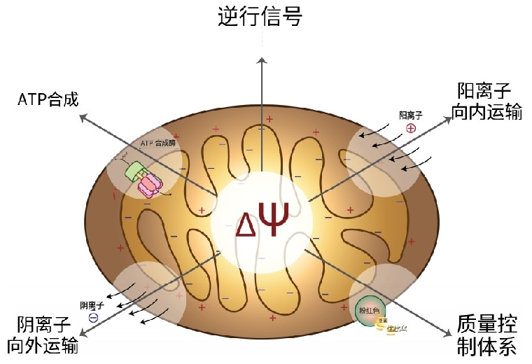
2. Mitochondrial membrane potential (MMP, ΔΨm) refers to the electrochemical potential formed by asymmetric distribution of protons and other ions across the inner mitochondrial membrane during cellular oxidative respiration. Intact MMP is critical for mitochondrial oxidative phosphorylation and ATP synthesis. Decrease or loss of MMP is associated with apoptosis, autophagy, necrosis and other cellular processes. Therefore, it serves as a key indicator for evaluating mitochondrial function and cell health. Detection of MMP changes helps reveal cellular energy metabolism and potential pathological alterations.
Figure 2: Mitochondrial membrane potential
3. JC‑1 used in this experiment is an ideal fluorescent probe widely applied for mitochondrial membrane potential measurement. As one of the lipophilic cationic compounds, JC‑1 selectively accumulates in the mitochondrial matrix and displays distinct fluorescence properties under different membrane‑potential levels.
4. In this experiment, users detect MMP levels by observing differential red‑green fluorescence intensity of JC‑1 within MDA‑MB‑231 cells, so as to judge cell apoptosis and provide experimental evidence for breast cancer research.
- Mitochondrial Membrane Potential Detection on MDA‑MB‑231 Cells
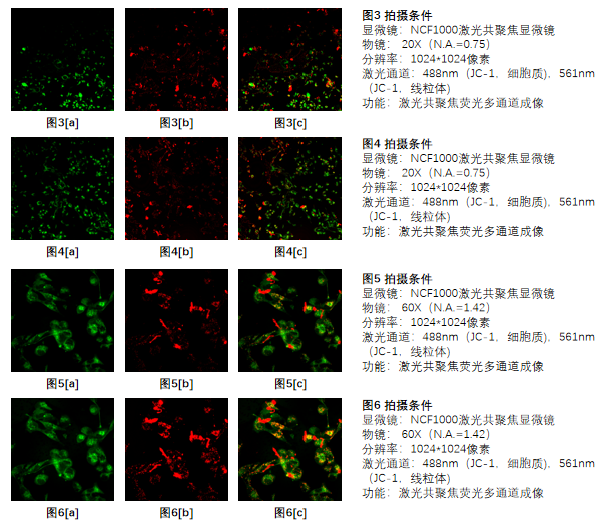
Within reasonable experimental conditions, normally‑growing MDA‑MB‑231 cells maintain high MMP, where JC‑1 forms aggregates with dominant red fluorescence. In apoptotic MDA‑MB‑231 cells, MMP declines and JC‑1 remains in monomer form with dominant green fluorescence.
Figure 3‑6: MDA‑MB‑231 cells
In this experiment, 20× and 60× objectives were used to observe intracellular fluorescence intensity of MDA‑MB‑231 cells. MMP levels were analyzed for preliminary assessment of cell viability. Comparing Figure 3 and Figure 4: under 20× objective, green fluorescence outweighs red fluorescence, indicating low MMP and overall apoptotic status, with more obvious apoptosis in Figure 4. Comparing Figure 5 and Figure 6: green fluorescence is more prominent under 60× objective, showing severe apoptosis; cells in Figure 5 are in worse condition than those in Figure 6. MMP measurement of MDA‑MB‑231 cells from different experimental groups provides experimental support for breast‑cancer therapy research.
II. How the NCF1000 Confocal Microscope Supports the Experiment
1. Simultaneous / Time‑sequential Multi‑channel Fluorescence Scanning
- The NCF1000 laser confocal microscope is equipped with four fluorescence photomultiplier tubes. The software supports simultaneous or time‑sequential scanning for up to four fluorescence channels (Figure 6).
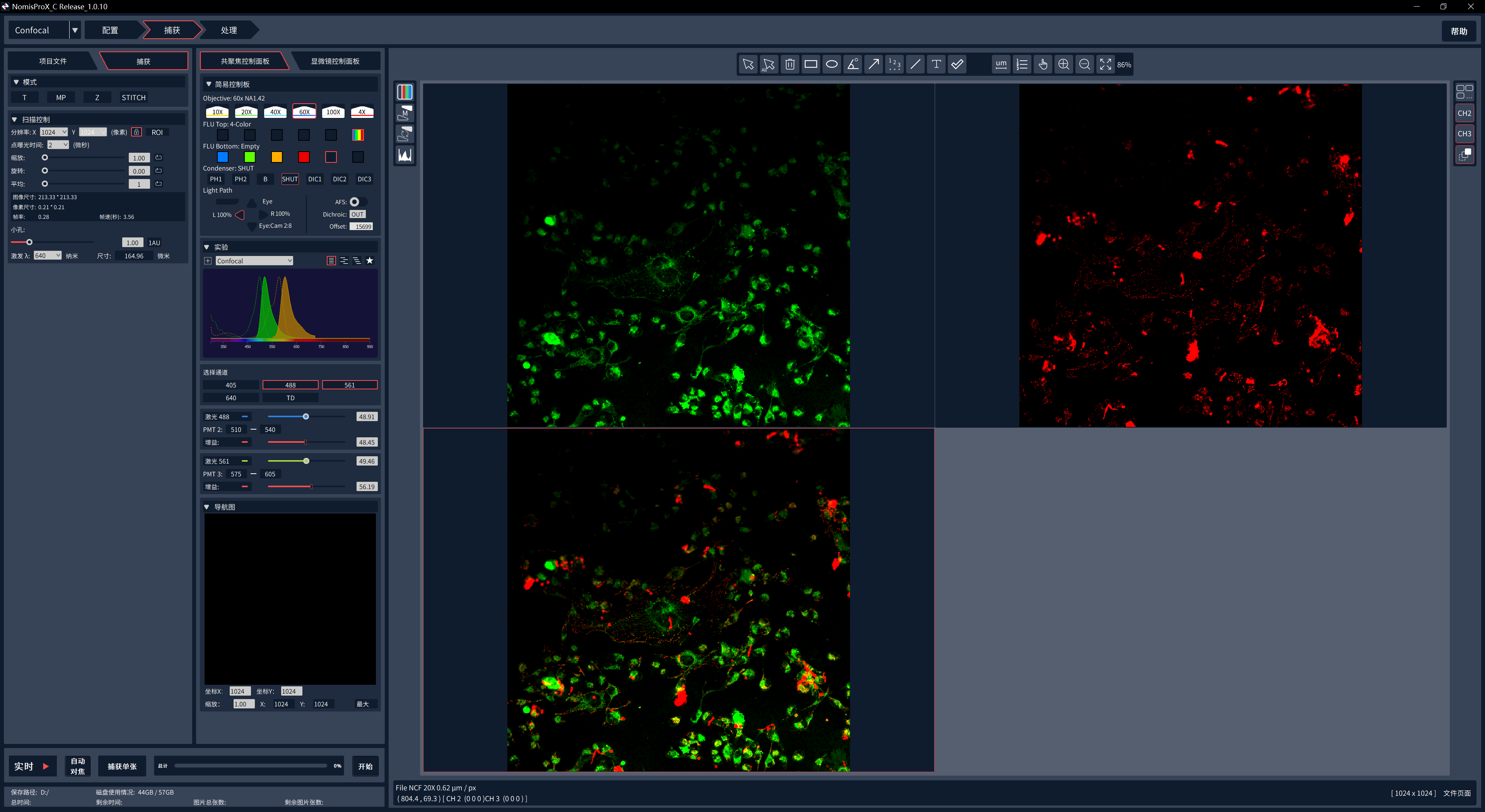
Figure 6: Multi‑channel fluorescence real‑time imaging interface
III. Common Experimental Issues
Q1: How to quickly locate the focal plane with 60× or higher‑magnification objectives?
A1: The NCF1000 laser confocal microscope provides three Z‑axis focusing methods. ① The focusing knobs on both sides of the microscope main unit offer low‑/medium‑/high‑speed gears. Locate the approximate focal plane at medium speed, then switch to low speed for fine adjustment. ③ Three‑speed Z‑axis focusing is also available via the motorized joystick following the same workflow above. ③ Click “Microscope Control Panel” in the software to customize focusing precision, then fine‑tune the focal plane with mouse wheel or Z‑axis movement.
Q2: How to avoid blurry mitochondrial structures when using 60× or higher‑magnification objectives?
A2: The NCF1000 laser confocal microscope provides three Z‑axis focusing methods. ① The focusing knobs on both sides of the microscope main unit offer low‑/medium‑/high‑speed gears. Locate the approximate focal plane at medium speed, then switch to low speed for fine adjustment. ② Three‑speed Z‑axis focusing is also available via the motorized joystick following the same workflow above. ③ Click “Microscope Control Panel” → “Stage Control” in the software to customize focusing precision, then fine‑tune the focal plane with mouse wheel or Z‑axis movement.

 Products